


While many skin lumps turn out to be harmless fatty tumors or benign growths, others require prompt veterinary attention. One of the most common malignant skin tumors seen in dogs is the mast cell tumor (MCT).
Mast cell tumors are often referred to as "the great pretenders" because they can look like almost any type of skin lump. Some appear as small, soft bumps that seem harmless, while others become swollen, ulcerated, or rapidly increase in size. What makes these tumors particularly challenging is that their appearance alone cannot determine whether they are benign or aggressive (Misdrop, 2004).
The good news is that advances in veterinary medicine have significantly improved the diagnosis and treatment of mast cell tumors. Many dogs diagnosed early go on to live happy, healthy lives, especially when tumors are removed before they have a chance to spread.
This guide explains everything pet owners need to know about mast cell tumors in dogs, including risk factors, symptoms, diagnosis, and what to expect if your dog receives this diagnosis.
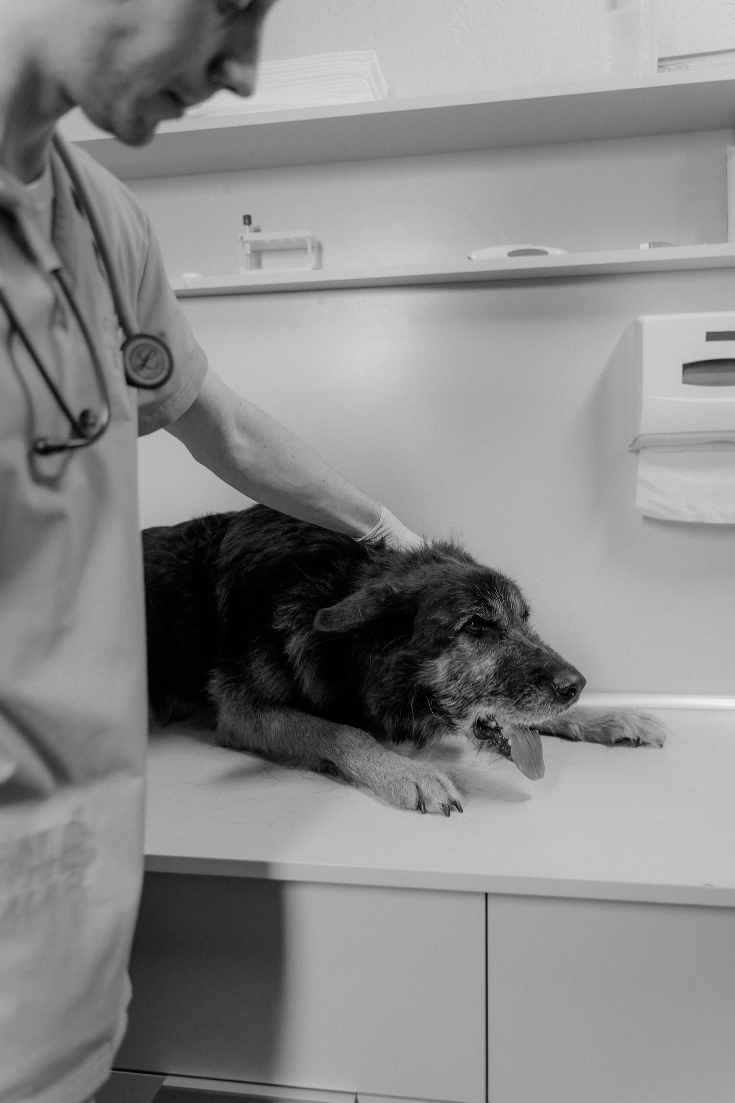
What Are Mast Cell Tumors?
Mast cell tumors develop from mast cells, a normal type of white blood cell and immune cell involved in allergic responses that can become cancerous. These specialized cells are found throughout the body but are especially concentrated in the skin, respiratory tract, and digestive system (Misdrop, 2004).
Under normal circumstances, mast cells help defend the body against parasites and participate in allergic reactions. They contain tiny granules filled with powerful chemicals, like histamine, heparin, enzymes, immune mediators and other inflammatory mediators.
When activated, mast cells release these substances to fight infection or respond to allergens.
Problems arise when these cells begin multiplying uncontrollably. Instead of functioning normally, they form tumors that can invade surrounding tissues and, in some cases, spread to lymph nodes, the spleen, liver, bone marrow, or other organs.
Mast cell tumors account for approximately 16–21% of all canine skin tumors, making them the most common skin tumor in dogs and a common skin tumor seen as nodules or masses in the skin.
Why Are Mast Cell Tumors So Unique?
Unlike many other cancers, mast cell tumors can change dramatically from day to day.
A tumor may appear small one morning and become noticeably larger by evening. Then, just as suddenly, it may shrink again.
This happens because histamine release and other inflammatory chemicals from the cancerous mast cells spill into the surrounding tissues. The swelling and irritation result from the substances involved in mast cell granules. These chemicals cause swelling, redness, itching, and inflammation (Horta et al., 2018).
This "waxing and waning" appearance is one of the hallmarks of mast cell tumors and often confuses pet owners into believing the lump is improving when it actually requires veterinary evaluation.
Additionally, manipulating the tumor (like squeezing or excessive touching) can trigger mast cell degranulation. This can release more histamine, temporarily making the mass appear larger or more irritated.
What Causes Mast Cell Tumors?
The exact cause remains unknown, but researchers believe multiple factors contribute to their development.
Genetics
Certain breeds have a significantly higher risk, suggesting a strong genetic component.
In some dogs, mutations affecting cell growth (especially involving the KIT gene) allow mast cells to multiply uncontrollably. These genetic changes may also influence mast cell tumor development. These mutations are especially common in more aggressive tumors.
Age
Most mast cell tumors occur in middle aged to older dogs between 8 and 10 years of age.
However, younger dogs are not immune, and some aggressive tumors develop in dogs as young as three or four years old.
Chronic Inflammation
Although evidence is still emerging, chronic skin inflammation and repeated irritation may contribute to abnormal mast cell activity in susceptible dogs.
Environmental Factors
Unlike many human cancers, no single environmental toxin has been conclusively linked to mast cell tumors in dogs.
Researchers continue studying possible influences, but genetics appears to play the largest role.

Which Dogs Are Most at Risk?
Although any breed can develop mast cell tumors, certain breeds have a much higher incidence.
These include Boxers, Labrador Retrievers, Golden Retrievers, Boston Terriers, Pugs, Staffordshire Bull Terriers, English Bulldogs, French Bulldogs, Weimaraners, Rhodesian Ridgebacks, Beagles, Schnauzers, and Shar Peis. Interestingly, different breeds tend to develop different types of tumors.
For example:
-
Boxers frequently develop lower-grade tumors that often respond well to surgery.
-
Shar-Peis are unfortunately predisposed to highly aggressive mast cell tumors at a younger age.
-
Golden Retrievers commonly develop multiple mast cell tumors during their lifetime.
Mixed breed dogs can also develop mast cell tumors, so no dog should be considered completely risk free.
Where Do Mast Cell Tumors Develop?
Around 80% of canine mast cell tumors occur within the skin, where they present as a cutaneous mast cell tumor.
Common locations include legs, chest, neck, flanks, abdomen, face and tail. However, they can develop almost anywhere on the body.
Less commonly, mast cell tumors form internally, affecting organs such as spleen, liver, gastrointestinal tract, and bone marrow. Internal mast cell tumors are considerably more serious because they are often diagnosed later in the disease process.
Signs in your pet
One reason mast cell tumors are nicknamed "the great imitator" is that the clinical signs rarely look the same from one dog to another.
Possible appearances include:
-
A small button-like bump
-
A hairless raised lump
-
A soft swelling beneath the skin
-
A wart like growth
-
A red, inflamed mass
-
An ulcerated lesion
-
A rapidly growing nodule
Some tumors remain unchanged for months, while others double in size within days.
Additional signs may include itching, redness, swelling, bleeding, scabbing, pain when touched and hair loss over the lump.
Because mast cells release histamine, some dogs develop skin changes and whole-body symptoms after histamine release from the tumor, especially if the tumor is aggressive.
These may include vomiting, diarrhea, loss of appetite, stomach ulcers, black/tarry stools, lethargy and weight loss.
These gastrointestinal signs occur because histamine stimulates excessive stomach acid production, which can lead to ulcer formation, and black, tarry stools are called melena.
Why You Should Never Ignore a Lump
Many owners assume that a lump is "just a fatty tumor." Unfortunately, mast cell tumors can closely resemble benign lipomas. Veterinarians often emphasize one simple rule: Every new lump deserves evaluation.
Even a suspicious skin tumor that appears harmless can be malignant. Fortunately, obtaining a diagnosis is usually quick and minimally invasive.
How Veterinarians Diagnose Mast Cell Tumors
The first and most important diagnostic tool is a fine needle aspiration (FNA).
This simple procedure involves inserting a small needle into the lump to collect cells.
Unlike surgical biopsies, most FNAs:
-
Require little or no sedation
-
Cause minimal discomfort
-
Can often be performed during a routine office visit
-
Provide results within one to three days
Under the microscope, mast cells are usually easy for pathologists to recognize because they contain characteristic purple staining granules. However, occasionally the cells are poorly differentiated, requiring additional testing.
If mast cells are identified, your veterinarian may recommend other diagnostic tests, including surgical biopsy, histopathology, blood work, lymph node aspiration, abdominal ultrasound, chest imaging, and other advanced staging tests. These additional evaluations help determine whether the cancer has spread and guide treatment planning (Garrett, 2014).

The Importance of Early Detection
Timing can make an enormous difference in outcomes.
Small tumors that are completely removed before spreading often carry an excellent prognosis.
Waiting several weeks or months to monitor a suspicious lump may allow a tumor to become larger, invade deeper tissues, or metastasize to distant organs. Routine monthly "nose-to-tail" examinations at home can help owners identify new lumps early. During grooming or cuddle time, gently run your hands over your dog's entire body and schedule a veterinary visit if you discover any new growth, especially one that changes in size, color, or texture. Remember: not every lump is cancer, but every lump deserves attention.
Grading and Staging: Understanding How Serious the Tumor Is
After a mast cell tumor has been diagnosed, the next step is determining how aggressive it is and whether it has spread. Veterinarians do this through grading and staging (Garrett, 2014).
Although the terms are sometimes used interchangeably, they describe two different aspects of the disease.
-
Grade refers to how abnormal the cancer cells look under a microscope and how aggressively they are expected to behave.
-
Stage refers to how far the cancer has spread throughout the body.
Understanding both helps your veterinarian recommend the most appropriate treatment plan and estimate your dog's prognosis.
Tumor Grading
A veterinary pathologist examines the tumor after it has been surgically removed or biopsied, using the submitted tissue sample for review. The cells are evaluated based on how organized they appear, how quickly they are dividing, and how deeply they invade surrounding tissues. Grading and related markers help predict prognosis and more accurately predict biological behavior.
The older Patnaik grading system classifies mast cell tumors into three grades:
Grade I (Low Grade)
These tumors are well-differentiated, meaning the cells still resemble normal mast cells. They are usually confined to the skin, grow slowly, and rarely spread. Surgical removal alone is often curative. Median survival time is often more than two years.
Grade II (Intermediate Grade)
These tumors vary considerably in behavior. Some behave like low grade tumors, while others are much more aggressive. Because of this unpredictability, Grade II tumors may require additional testing and close monitoring.
Grade III (High Grade)
These poorly differentiated tumors are aggressive, invasive, and much more likely to metastasize. Dogs with Grade III tumors often require surgery combined with chemotherapy, radiation therapy, or targeted medications. Average survival time is often less than four months.
Many veterinary pathologists now use the Kiupel two tier grading system, which classifies tumors as either low grade or high grade. In selected cases, an MCT prognostic panel may be recommended after diagnosis. This newer system is considered more consistent and helps veterinarians better predict outcomes.
Staging the Disease
Once the tumor has been graded, your veterinarian may recommend staging tests to check whether your dog's tumor has spread, such as to regional lymph nodes or as enlarged lymph nodes on exam.
Depending on your dog's condition, these tests may include:
-
Complete blood count (CBC)
-
Blood chemistry panel
-
Urinalysis
-
Fine needle aspirate of regional lymph nodes
-
Abdominal ultrasound
-
chest X-rays
-
CT scan (in some cases)
-
Bone marrow evaluation for advanced disease
Fortunately, many mast cell tumors are discovered before widespread metastasis occurs, and staging results can help determine whether further treatment is needed.
Treatment Options
Mast cell tumors treated based on several factors, including:
-
Tumor grade
-
Tumor location
-
Tumor size
-
Whether the cancer has spread
-
Your dog's age and overall health
Fortunately, there are several effective treatment options available.
Surgery
Surgical removal remains the gold standard treatment for most mast cell tumors.
The goal is to remove not only the visible tumor but also a margin of healthy tissue surrounding it. Mast cell tumors often extend microscopic "roots" beyond what is visible to the naked eye (Sledge et al., 2016).
Historically, surgeons aimed for 2–3 cm margins whenever possible, although newer studies suggest that smaller margins may be sufficient for many low-grade tumors located in favorable areas.
After surgery, the tissue is sent to a laboratory to determine whether the margins are "clean," meaning no cancer cells are present at the edges of the specimen.
Clean margins significantly reduce the risk of recurrence.
Radiation Therapy
Radiation therapy is commonly recommended when:
-
Complete surgical removal isn't possible.
-
Tumor cells remain at the surgical margins.
-
The tumor is located in an area where wide surgical margins cannot be achieved, such as the face or lower limbs.
Modern radiation therapy is generally well tolerated by dogs. Most continue eating, playing, and enjoying a good quality of life throughout treatment.
Chemotherapy
Chemotherapy is often recommended for:
-
High grade tumors
-
Multiple mast cell tumors
-
Tumors that have spread
-
Tumors with high-risk characteristics
Unlike human chemotherapy, veterinary chemotherapy is designed to preserve quality of life. Most dogs experience few serious side effects.
Common medications include:
-
Vinblastine
-
Prednisone
-
Lomustine (CCNU)
Possible side effects include mild gastrointestinal upset, temporary loss of appetite, lower white blood cell counts, and fatigue (De Nardi et al., 2022).
Hair loss is uncommon in dogs, although breeds with continuously growing coats, such as Poodles and Old English Sheepdogs, may experience some thinning.
Targeted Therapy
One of the most exciting advances in veterinary oncology is the development of targeted therapies known as tyrosine kinase inhibitors (TKIs).
Some mast cell tumors contain mutations in the KIT gene, which causes uncontrolled cell growth.
Medications such as:
-
Toceranib phosphate (Palladia®)
-
Masitinib (Hahn et al., 2008)
block these abnormal signaling pathways and may slow tumor growth in selected dogs.
These medications are often used when surgery is not possible or when tumors recur after treatment.
Antihistamines and Supportive Care
Because mast cell tumors release large amounts of histamine, many dogs receive medications to minimize the effects of this chemical (De Nardi et al., 2022).
Your veterinarian may prescribe:
-
Diphenhydramine (Benadryl®)
-
Famotidine
-
Omeprazole
-
Sucralfate
These medications help reduce:
-
Allergic reactions
-
Stomach ulcers
-
Vomiting
-
Gastrointestinal bleeding
Supportive care plays an important role in keeping dogs comfortable throughout treatment (Sledge et al., 2016).

Prognosis
One of the first questions owners ask is:
"Will my dog survive?"
The answer depends on several factors.
Dogs with completely removed low grade mast cell tumors often have an excellent prognosis and may never experience another problem.
Many live normal life spans without recurrence.
High grade tumors carry a more guarded prognosis because they are more likely to spread to internal organs (Thompson et al., 2011).
Factors associated with a better outcome include low tumor grade, clean surgical margins, small tumor size, no lymph node involvement, no evidence of metastasis, early diagnosis (Thompson et al., 2011).
Even dogs with more advanced disease may respond well to modern treatment combinations, allowing many to enjoy months or even years of good quality of life (Romansik et al., 2007).
Can Mast Cell Tumors Be Prevented?
Unfortunately, there is no guaranteed way to prevent mast cell tumors. Because genetics plays such a significant role, prevention is not always possible. However, early detection remains the best strategy for improving outcomes.
Pet owners should perform monthly skin checks, schedule annual or twice yearly veterinary examinations, have every new lump evaluated promptly, never assume a lump is "just a fatty tumor," and monitor existing masses for changes in size, shape, or color.
Maintaining a healthy weight, feeding a balanced diet, and keeping up with routine veterinary care can also support your dog's overall health, although these measures have not been proven to prevent mast cell tumors specifically.
Living with a Dog Diagnosed with a Mast Cell Tumor
Receiving a cancer diagnosis can be overwhelming, but many owners are surprised to learn that mast cell tumors are often treatable.
Dogs do not dwell on their diagnosis the way humans do. They focus on enjoying each day; whether that means chasing a tennis ball, going for walks, or relaxing beside their favorite person.
Working closely with your veterinarian or a board-certified veterinary oncologist can help you make informed decisions about treatment while prioritizing your dog's comfort and quality of life.
Many families report that, after successful treatment, their dogs return to their normal routines and continue to enjoy life for years.
Final Thoughts
Mast cell tumors are among the most common skin cancers diagnosed in dogs, but they are also among the most manageable when detected early. Because these tumors can mimic harmless skin lumps, every new bump should be evaluated by a veterinarian using a fine needle aspirate or other appropriate diagnostic tests.
Advances in surgery, radiation therapy, chemotherapy, and targeted medications have dramatically improved outcomes for many dogs. While some mast cell tumors behave aggressively, many low-grade tumors can be successfully treated with surgery alone, allowing dogs to live long, happy, and active lives.
The most important takeaway for pet owners is simple: don't ignore a new lump. Early diagnosis offers the best chance for successful treatment and can make a life-changing difference for your canine companion. If you ever notice a new bump, swelling, or skin growth on your dog, schedule a veterinary examination promptly. Acting quickly could give your pet the best possible outcome.
References
De Nardi, A. B., dos Santos Horta, R., Fonseca-Alves, C. E., de Paiva, F. N., Linhares, L. C. M., Firmo, B. F., ... & Dagli, M. L. Z. (2022). Diagnosis, prognosis and treatment of canine cutaneous and subcutaneous mast cell tumors. Cells, 11(4), 618.
Garrett, L. D. (2014). Canine mast cell tumors: diagnosis, treatment, and prognosis. Veterinary Medicine: Research and Reports, 49-58.
Hahn, K. A., Oglivie, G., Rusk, T., Devauchelle, P., Leblanc, A., Legendre, A., ... & Hermine, O. (2008). Masitinib is safe and effective for the treatment of canine mast cell tumors. Journal of Veterinary Internal Medicine, 22(6), 1301-1309.
Horta, R. S., Lavalle, G. E., Monteiro, L. N., Souza, M. C., Cassali, G. D., & Araújo, R. B. (2018). Assessment of canine mast cell tumor mortality risk based on clinical, histologic, immunohistochemical, and molecular features. Veterinary pathology, 55(2), 212-223.
Misdorp, W. (2004). Mast cells and canine mast cell tumours. A review. Veterinary quarterly, 26(4), 156-169.
Romansik, E. M., Reilly, C. M., Kass, P. H., Moore, P. F., & London, C. A. (2007). Mitotic index is predictive for survival for canine cutaneous mast cell tumors. Veterinary pathology, 44(3), 335-341.
Sledge, D. G., Webster, J., & Kiupel, M. (2016). Canine cutaneous mast cell tumors: A combined clinical and pathologic approach to diagnosis, prognosis, and treatment selection. The Veterinary Journal, 215, 43-54.
Thompson, J. J., Pearl, D. L., Yager, J. A., Best, S. J., Coomber, B. L., & Foster, R. A. (2011). Canine subcutaneous mast cell tumor: characterization and prognostic indices. Veterinary pathology, 48(1), 156-168.
Related Posts

Diabetes in Dogs and Cats
Diabetes mellitus is a common endocrine disease in dogs and cats. While a diabetes diagnosis can initially feel overwhelming, many diabetic pets can continue to enjoy a good quality of life with appropriate veterinary care and consistent...

How to compare mushroom supplements: Buddy Guard vs. Critical Immune Defense
This comparison is published by Petsmont, the maker of Buddy Guard on August 5th, 2026. Competitor information came from publicly available labels and manufacturer materials. TLDR: Summary Table Buddy Guard Critical Immune Defense ...

The Hyperthyroid Cat: A Complete Guide for Pet Owners
As cats grow older, they become more susceptible to certain age-related health conditions. Hyperthyroidism in cats is usually one of the most common endocrine disorders in senior cats. This is an overproduction of thyroid hormones that s...


Leave a comment